|
Home / Common_In_Lake / Attitash / 20160908 / Images |
|
Attitash 20160908 (Amesbury, MA) |
|
|
|
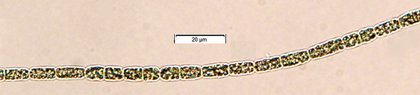
Anabaena spp. Three 'species' or strains with different cell size and shape. Horizontal filament in lower image has a hyaline sheath, unusual in the genus, as well as an akinete (spore) on another trichome - the larger greener cell.
|
|
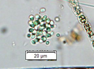
Aphanocapsa sp.
|
|
Ceratium hirundinella exploded under cover slip
|
|
Cyclotella cf. comta or meneghiniana valve
|
|
Euglena sp.
|
|
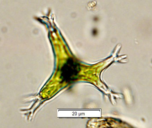
Isthmochloron sp. (Tribophyceae)
|
|
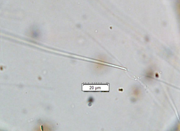
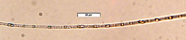
Lyngbya cf. pusilla, the
narrowest filament in this genus
|
|
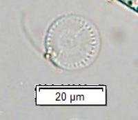
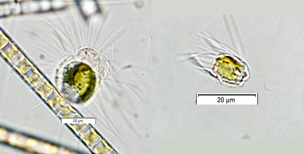
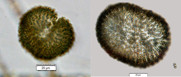
Mallomonas sp.
|
|
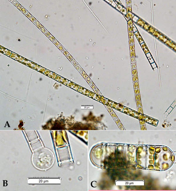
Melosira sp. showing (A) vegetative trichome (filament lacking sheath); (B) developing auxospore; (C) mature auxospore with >2x diameter of vegetative cells.
|
|
Microcystis cf. aeruginosa Joosten, A.M.T. (2006). Flora of the blue-green algae of the Netherlands I The non-filamentous species of inland waters. pp. [1-]5-237. Utrecht: KNNV Publishing.
|
|
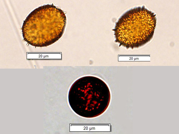
Trachelomonas spp.
|
|
Woronochinia sp.
|
|
Oscillatoria
cf. redekii
|
|
Home / Common_In_Lake / Attitash / 20160908 / Images |