|
Home / Non-PS_Flagellates_and_Ciliates / Ciliates / Dictyocysta / Images |
|
Dictyocysta
(Testate ciliate) |
|
|
|
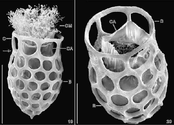
Dictyocysta mitra.
A ‘closing apparatus’ (Agatha 2010) activated by outside disturbance
can be seen in the right-hand image as the top of a membrane sack surrounding
cell and inside the lorica. SEM
photograph by © Sabine Agatha, Department
of Organismic Biology, University of Salzburg, Hellbrunnerstrasse
34, A‐5020
Salzburg, Austria, posted online. Agatha,
S. 2010. A light and scanning electron microscopic
study of the closing apparatus in tintinnid ciliates (Ciliophora,
Spirotricha, Tintinnina): A forgotten synapomorphy. Eukaryotic Microbiology 57(4):297-307. Agatha, S. 2010. A forgotten
synapomorphy in tintinnid ciliates (Ciliophora, Spirotricha, Tintinnina): a
light and scanning electron microscopic study of the closing apparatus.
Science in Parasitology and Protozoology solves Problems. Düsseldorf,
Germany.
|
|
Home / Non-PS_Flagellates_and_Ciliates
/ Ciliates / Dictyocysta / Images |