| Gymnodinium (Dinophyceae) |
||||
|
|
||||
|
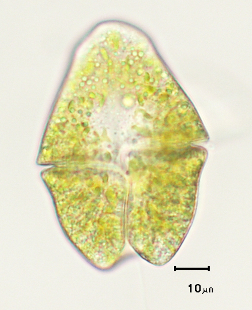
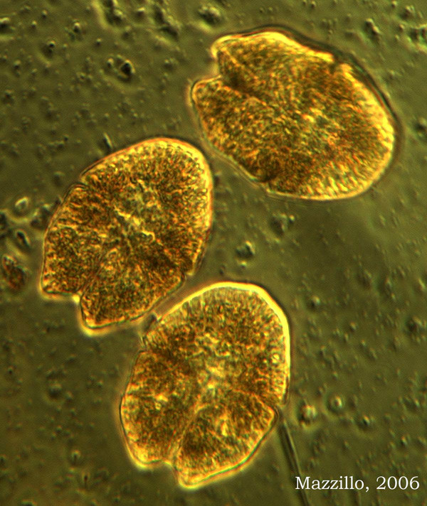
Two flagella, one trailing and the other wrapped around the equatorial groove, are not visible in these images.
|
||||
|
|
||||
|
|
||||
|
|
||||
|
|
||||
|
|
||||
|
|
||||
|
|
||||
Image shows thin cellulose cell wall separating .
|
||||
Lack of heavy cellulosic plates is evident in this image. |
||||
Gymnodinium sanguinea |
||||
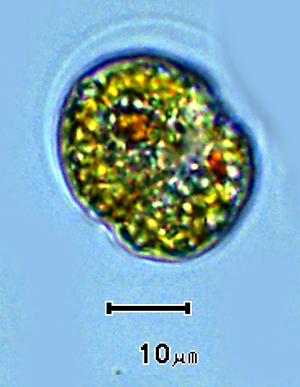
Gymnodinium sp. in palmoloid stage - loss of flagella, gain of thick mucilaginous sheath.
|
||||
|
|
||||
|
Home / Dinoflagellates /Photosynthetic / Gymnodinium / Images |
||||