|
Home / Synurophyceae / Mallomonas / Images |
|
|
|
Mallomonas (Synurophyceae) |
|
|
|
|
|
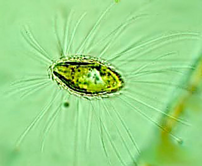
Mallomonas sp.
|
|
|
|
Mallomonas sp.
|
|
|
|
Mallomonas sp.
|
|
|
|
Mallomonas sp. from Dollar Bay,
Keweenaw Waterway, Lake Michigan USA
|
|
|
Mallomonas sp., from Wheelwright Pond, Lee, NH USA (2 July 2020)
|
|
|
|
Mallomonas sp., numerous silicaceous scales and bristles
|
|
|
|
Mallomonas sp., bristles dehydrated, cell still within water
|
|
|
|
Mallomonas sp., dehydrated cell emphasizing 5 - 6 teeth near bristle distal ends
|
|
|
|
Mallomonas sp., dehydrated cell emphasizing bristles
|
|
|
|
Mallomonas sp. scales, bristles, and
anterior flagellum, from Witten Hammerteich DE
|
|
|
|
SEM of Mallomonas cf. crassisquama illustrating bristle hooked 'feet' tucked underneath each scale. Photograph posted online
|
|
|
|
Mallomonas bakeri illustrating overlap of silicaceous scales Siver, P.A. 2018. Mallomonas skogstadii sp. nov. and M. bakeri sp. nov.: Two New Fossil Species from the Middle Eocene Representing Extinct Members of the Section Heterospinae?. Cryptogamie, Algologie 39(4), 511-524, https://doi.org/10.7872/crya/v39.iss4.2018.511
|
|
|
|
Mallomonas transsylvanica scale (scale bar 2 um)
|
|
|
|
Mallomonas insignis with rhomboid silicaceous
scales and caudal peduncle scales (arrowheads)
|
|
|
|
Mallomonas
caudatus bristles with sawtooth distal ends
|
|
|
Mallomonas silica spine from Rybníčky u Podbořánek nature reserve, Czech Republic,
illustrating unilaterally toothed distal end, shaft, and right-angle hooked
proximal end that is inserted into sheath of a flattened body scale extracellularly, allowing the spine to flex during
start-stop movements of flagella. Electron microscope photograph by Martin Pusztai posted online. |
|
|
|
|
|
Home / Synurophyceae / Mallomonas / Images |


